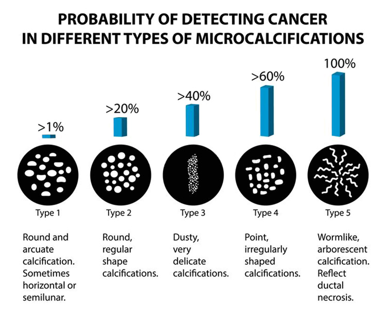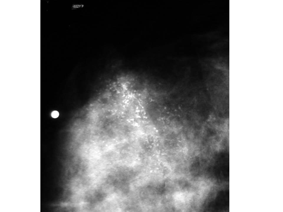

They may be punctate, linear, spherical, coarse, cylindrical, smooth, jagged, regular, casting, branching or heterogeneous, with the last three being the most alarming ones. SHAPE – The shape of the microcalcifications is probably the most important element in their analysis (fig. Shape – Size – Density – Number – Distribution – Location – Associated findings. Regardless of the final diagnosis, calcifications should be assessed according to well-established attributes. There are calcifications whose characteristics denote a benign process and other calcifications whose characteristics hint a malignant one. When attached to an otherwise “benign-looking” finding, they may reveal, through their characteristics, the true malignant nature of the whole process and on the contrary, by their benign attributes, an otherwise “suspicious-looking” finding may get a less invasive work-up. However, calcifications may be a sign of benign changes they may also disclose a yet nonpalpable malignant process.

While attempting to analyse breast calcifications, their characteristics have to be taken into account, combinations of these characteristics leading finally to a safe management of a given case. While dealing with breast calcifications, BIRADS takes into account distribution of the calcifications within a breast and patterns of calcifications, dealing less with basic morphological characteristics of calcifications. In order to standardize mammographic reporting, reduce confusing breast imaging interpretations and facilitate outcome monitoring, the American College of Radiology implemented the Breast Imaging Reporting and Database System, known as BIRADS (). Microcalcifications are best-visualized using high-resolution imaging techniques, vigorous compression and radiographic magnification. Some studies attempted to connect certain compositions or individual elements within calcifications to malignant or benign changes, in order to allow in vivo spectroscopy differentiate malignant from benign breast changes. Apatite, calcite, calcium oxalate, oxalic acid, aragonite and traces of aluminium, iron, magnesium, silicon, copper, gold, silver, titanium and some other elements have been identified. Their exact composition remains uncertain.
#BREAST CALCIFICATION CLUSTERS SKIN#
They may be located anywhere in the breast structures, including skin and interstitial stroma.Īctive cell secretion, cellular necrotic debris, inflammation, foreign body reaction, trauma and radiation are the most usual reasons for their formation. CALCIFICATIONS OF BREAST are the smallest structures identified on a mammogram and are always a sign of a past or ongoing breast tissue alteration.Įxtremely common, seen in up to 86% of the mammograms, they are usually benign and their frequency increases with age.


 0 kommentar(er)
